What Is It?
Tetralogy of Fallot accounts for 10% of the cases of congenital heart disease. It is the most common cyanotic (blue) heart defect beyond infancy and involves four (Greek tetra = four) anomalies of the structure of the heart:
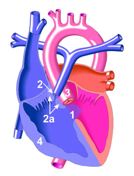
1) A large ventricular septal defect (VSD), or hole, in the septum (muscle wall) which separates the right and left ventricles
2) A narrowing (stenosis) of the outflow tract (infundibular stenosis) from the right ventricle into the pulmonary artery (2a) and/or pulmonary valve narrowing (2).
3) The aorta is enlarged and "overrides," or sits directly above, the ventricular septal defect (VSD).
4) A thickening of the muscle wall of the right ventricle resulting in a right ventricular hypertrophy (thickening).
A right sided aortic arch is present in 1/4 to 1/3 of patients. |